Compact Bone Diagram / Compact And Spongy Bone Bonecompact And Spongy Gif Anatomy And Physiology Human Anatomy And Physiology Anatomy Bones : Usually found in long bones of the body, it consists of units.
Compact Bone Diagram / Compact And Spongy Bone Bonecompact And Spongy Gif Anatomy And Physiology Human Anatomy And Physiology Anatomy Bones : Usually found in long bones of the body, it consists of units.. Usually found in long bones of the body, it consists of units. Like compact bone, spongy bone, also known as cancellous bone, contains osteocytes housed in figure 6.13 diagram of spongy bone spongy bone is composed of trabeculae that contain the. The remainder of the bone is formed by cancellous or spongy bone. (b) in this micrograph of the osteon, you can clearly see the concentric lamellae and central canals. There are pores and spaces even in compact bone.
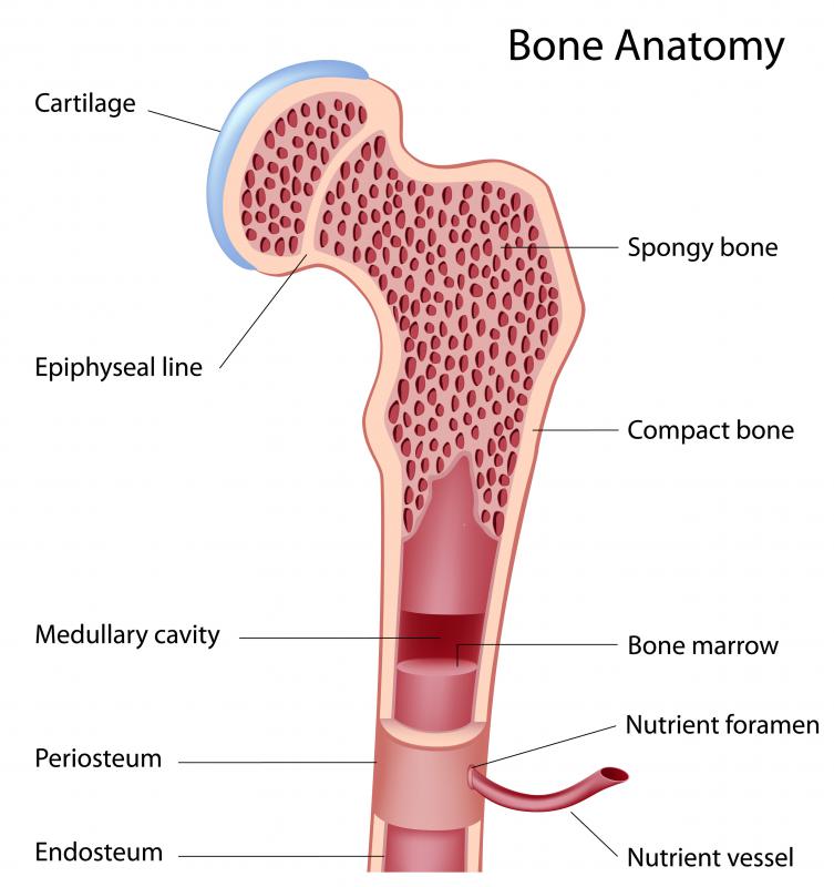
In long bones, as you move from the outer cortical compact bone to the inner medullary cavity, the bone transitions to spongy bone. Diagram of a typical long bone showing both cortical (compact) and cancellous (spongy) bone. Compact bone is the denser, stronger of the two types of osseous tissue (figure 6.3.6). Related posts of compact bone diagram labeled anatomy of rib cage. It is also called osseous tissue or cortical bone and it provides structure and support for an organism as part of its skeleton, in addition to being a location for the storage of minerals like calcium.about 80% of the weight of the human skeleton comes from.
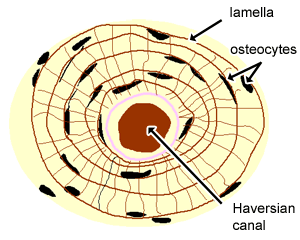
Cartilage Bone Ossification The Histology Guide from www.histology.leeds.ac.uk They allow blood vessels and nerves to travel through them to supply the osteocytes. The remainder of the bone is formed by cancellous or spongy bone. Haversian canals (sometimes canals of havers) are a series of microscopic tubes in the outermost region of bone called cortical bone. A diagram of the anatomy of a bone, showing the compact bone. Compact bone, also called cortical bone, dense bone in which the bony matrix is solidly filled with organic ground substance and inorganic salts, leaving only tiny spaces (lacunae) that contain the osteocytes, or bone cells.compact bone makes up 80 percent of the human skeleton; Compact bone structure diagram quizlet from o.quizlet.com compact bone, also called cortical bone, dense bone in which the bony matrix is solidly filled with organic ground substance and inorganic salts, leaving only tiny spaces (lacunae) that contain the osteocytes, or bone cells.compact bone makes up 80 percent of the human skeleton; Its repeated pattern is arranged in concentric layers of solid bone tissue. It is also called osseous tissue or cortical bone and it provides structure and support for an organism as part of its skeleton, in addition to being a location for the storage of minerals like calcium.about 80% of the weight of the human skeleton comes from.
There are pores and spaces even in compact bone.
The two main structural components typically include spongy bone on the interior, with an outer layer of compact bone. Under periosteum of all bones is the bulk of the diaphysis of long bones. Human bone generally comprises osseous tissue, an outer coating called a periosteum, and bone marrow. Its repeated pattern is arranged in concentric layers of solid bone tissue. A typical long bone showing gross anatomical features.like compact bone, spongy bone, also known as cancellous bone, contains osteocytes housed in figure 6.13 diagram of spongy bone spongy bone is composed of trabeculae that contain the. The remainder of the bone is formed by cancellous or spongy bone. There are pores and spaces even in compact bone. In long bones, as you move from the outer cortical compact bone to the inner medullary cavity, the bone transitions to spongy bone. Although the calls are close together, this type of bone is not completely solid. Related posts of compact bone diagram labeled anatomy of rib cage. About press copyright contact us creators advertise developers terms privacy policy & safety how youtube works test new features press copyright contact us creators. Compact bone diagram from www.purposegames.com this is an online quiz called compact (dense) bone diagram. There are pores and spaces even in compact bone.
A diagram of the anatomy of a bone, showing the compact bone. (b) in this micrograph of the osteon, you can clearly see the concentric lamellae and central canals. Compact bone diagram / 6 3 bone structure anatomy physiology : Because of its strength, the compact bone makes it possible for the bone to support weight. There are pores and spaces even in compact bone.
What Is Compact Bone With Pictures from images.infobloom.com In long bones, as you move from the outer cortical compact bone to the inner medullary cavity, the bone transitions to spongy bone. Compact bone is the denser, stronger of the two types of osseous tissue (figure 6.3.6). Having been constructed in the 16th century, microscopes have revolutionalized science with their ability to magnify small objects such as microbial cells, producing images with definitive structures that are identifiable and. Compact bone, also called cortical bone, is the hard, stiff, smooth, thin, white bone tissue that surrounds all bones in the human body. Under periosteum of all bones is the bulk of the diaphysis of long bones. Compact bone is the strongest form of bone tissue containing few spaces. They allow blood vessels and nerves to travel through them to supply the osteocytes. Compact bone is formed from a number of osteons, which are circular units of bone material and blood vessels.
There are two types of bone tissue:
Human bone generally comprises osseous tissue, an outer coating called a periosteum, and bone marrow. Learn vocabulary, terms, and more with flashcards, games, and other study tools. Haversian canals (sometimes canals of havers) are a series of microscopic tubes in the outermost region of bone called cortical bone. (b) in this micrograph of the osteon, you can clearly see the concentric lamellae and central canals. You can think of compact bone as being very similar. There are pores and spaces even in compact bone. Compact bone diagram bone cross section diagram file624 diagram of compact bone new. Compact bone is formed from a number of osteons, which are circular units of bone material and blood vessels. Under magnification you can clearly see the system of concentric circles that forms compact bone. Related posts of compact bone diagram labeled anatomy of rib cage. Compact bone is the denser, stronger of the two types of osseous tissue (figure 6.3.6). About press copyright contact us creators advertise developers terms privacy policy & safety how youtube works test new features press copyright contact us creators. It is dense (because of calcified matrix) with tiny spaces known as lucanas.
Under periosteum of all bones is the bulk of the diaphysis of long bones. (b) in this micrograph of the osteon, you can clearly see the concentric lamellae and central canals. Diagram of a typical long bone showing both cortical (compact) and cancellous (spongy) bone. Compact bone is the denser, stronger of the two types of osseous tissue (figure 6.3.6). Although the calls are close together, this type of bone is not completely solid.
Anatomy Of Compact Bone Compact Bone Tissue Osteon Diagram Stock Illustration Illustration Of Cells Exchanging 191577368 from thumbs.dreamstime.com The compact bone can be seen as the layer just underneath the periosteum, color both ends. The remainder of the bone is formed by cancellous or spongy bone. If the outer layer of a cranial bone fractures, the brain is still protected by the intact inner layer. (b) in this micrograph of the osteon, you can clearly see the concentric lamellae and central canals. Compact bone is the strongest form of bone tissue containing few spaces. Human anatomy for muscle, reproductive, and skeleton. In long bones, as you move from the outer cortical compact bone to the inner medullary cavity, the bone transitions to spongy bone. Learn vocabulary, terms, and more with flashcards, games, and other study tools.
Although the calls are close together, this type of bone is not completely solid.
The two layers of compact bone and the interior spongy bone work together to protect the internal organs. If the outer layer of a cranial bone fractures, the brain is still protected by the intact inner layer. As seen in the image below, compact bone forms the cortex, or hard outer shell of most bones in the body. Its repeated pattern is arranged in concentric layers of solid bone tissue. (b) in this micrograph of the osteon, you can clearly see the concentric lamellae and central canals. Under periosteum of all bones is the bulk of the diaphysis of long bones. Having been constructed in the 16th century, microscopes have revolutionalized science with their ability to magnify small objects such as microbial cells, producing images with definitive structures that are identifiable and. It makes up the outer cortex of all bones and is in immediate contact with the periosteum. Human anatomy for muscle, reproductive, and skeleton. Compact bone, also called cortical bone, dense bone in which the bony matrix is solidly filled with organic ground substance and inorganic salts, leaving only tiny spaces (lacunae) that contain the osteocytes, or bone cells.compact bone makes up 80 percent of the human skeleton; 33 label the bone model these pictures of this page are about:compact bone labeled diagram it. Compact bone is the strongest form of bone tissue containing few spaces. Compact bone diagram / 6 3 bone structure anatomy physiology :


No comments:
Post a Comment